摘要
膜翅目昆虫毒素富含数种致过敏物质, 如抗原-5蛋白、磷脂酶A、透明质酸酶和蛋白酶等, 其叮咬是引起速发型过敏反应的三大诱因之一(其他两类诱因为食物和药物), 并且膜翅目昆虫叮咬引起的过敏反应在人群中的发生率还在缓慢增加。叮咬后的常见症状有局部红肿和灼痛, 严重的可表现为过敏性休克、急性肾衰竭和多器官功能障碍综合征等. 目前对这类过敏反应尚无很好的治疗方法, 毒素过敏原特异性免疫疗法是治疗这类过敏反应的最有效手段。对这类过敏原的研究可为膜翅目昆虫叮咬引起过敏反应的诊治提供理论和物质基础。作为膜翅目昆虫的胡蜂, 其个体较大、毒素量丰富, 本文将结合本实验室近年来的研究工作, 对胡蜂毒素过敏原的组成成分及其引发过敏反应的作用机制作一简要综述。
关键词 胡蜂 毒素 过敏原 过敏反应机制
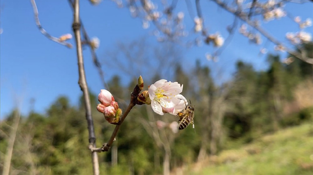
胡蜂(wasp), 亦名马蜂、黄蜂等, 隶属于膜翅目 (Hymenoptera)细腰亚目(Apocrita)胡蜂总科(Vespoidea)。 胡蜂种类较多, 目前世界已知胡蜂约6000余种, 我国已发现的胡蜂也达200 多种[1]. 胡蜂呈世界性的广泛分布, 胡蜂种属的命名常与地域有关, 不同属间胡蜂毒素组成成分差异较大[2]。胡蜂毒素引起的速发型过敏反应常常能造成人员的伤亡, 其人群发病率也在增加, 特别是在工业化国家[3]。美国罗切斯特流行病学项目的报告称, 膜翅目昆虫叮咬引起的速发型过敏反应人群发生率从20世纪80年代的十万分之二十一增至目前的十万分之五十[4]. 叮咬能引起过敏反应的常见膜翅目昆虫有蚊子、蜜蜂、胡蜂、牛虻等, 其中胡蜂和蜜蜂的毒素较为珍贵, 牛虻的唾液腺也较难获取, 它们在药理学、免疫学等领域具有重要的科研价值. 20世纪50年代, 国外科学家对胡蜂毒素的组成成分进行了初步的研究, 发现胡蜂毒素主要由胺、多肽、酶及其他未知功能的蛋白等组成[5]. 现代分子生物学技术的发展, 使得人们对蜂毒成分有了更加充分的认识. 属于胺类的组分有5-羟色胺、组胺、多巴胺和去甲肾上腺素等, 它们构成了胡蜂毒素小分子物质的主要成分[6]. 属于多肽类的有蜂毒肽 (melittin)、蜂毒明肽(apamin)、肥大细胞脱粒肽、缓激肽、抗菌肽、亲肌活性的毒素肽和化学趋化肽等[7~14]. 胡蜂毒素的蛋白电泳显示, 在23, 34和43 kD处的蛋白浓度较高, 经鉴定它们分别为抗原-5蛋白 (antigen-5 protein), 磷脂酶A1 (phospholipase A1)和透明质酸酶(hyaluronidase). 它们是胡蜂毒素中的3种主要蛋白成分, 也是胡蜂毒素导致过敏反应的主要 过敏原, 而且这3种蛋白与蜜蜂毒素、牛虻唾液腺的对应蛋白存在不同程度的交叉反应[15~18].
1 胡蜂毒素导致过敏的物质基础
1.1 抗原-5蛋白(antigen-5 protein, Ag5)
抗原-5是胡蜂毒素最主要过敏原之一, 最早由 King等人[19]报道. 此外, 在红火蚁(Solenopsis invicta)、黑火蚁(Solenopsis richteri)的毒素和牛虻(Tabanus yao)唾液腺中[18,20], Ag5 也是作为过敏原被发现的.目前在胡蜂总科(Vespoidea)的胡蜂属(Vespa) [21~23]、长黄胡蜂属(Dolichovepula) [24~27]、黄胡蜂属(Vespula) [28~30]和马蜂属(Polistes) [31~34]中共发现17种Ag5. 经序列比对发现, 来源于同一胡蜂属毒素中的Ag5相似性较高, 它们之间的相似性在87%~95%之间, 而来自不同属Ag5之间的相似性只有约60%, 这些 Ag5 之间都有不同程度的交叉免疫反应[35]. Ag5在毒素和唾液腺中的生物学功能目前尚不清楚[19]. 在 PROSITE 数据库中搜索Ag5, 发现Ag5与某些哺乳动物、爬行类、真菌和植物的一类蛋白具有较高相似性, 这类蛋白与发病机理有关或在再生组织、细胞中表达, 它们共同构成了一类蛋白超家族. 这类蛋白超家族的NMR显示它们的结构呈现一种独特的折叠方式, 这三明治结构,它们的核心……结构表现出一种折叠片构成……螺旋和4个……螺旋, 1个小……构是由3个大成[36]. 由于T淋巴细胞在过敏反应发生中起着接收抗原递呈细胞(antigen-presenting cell APC)递呈的过敏原多肽和帮助B淋巴细胞产生抗体等重要作用, Bohle 等人[37]对属于Ag5的Ves v 5的T细胞抗原表位(T cell epitopes)进行了研究. 利用合成的65条来自Ves v 5的重叠多肽和Ves v 5特异性的T细胞系, 从中鉴定了28个T 细胞抗原表位, 其中Ves v 5181~192 这段多肽是主要的T细胞抗原表位, 并且这段序列在胡蜂总科的各个Ag5中均较为保守. King 等人[35] 对Ag5 Ves v 5和Pol a 5进行重组, 表达出 9 种Ves v 5和Pol a 5的杂合体, 对Ag5的B细胞表位(B cell epitopes)进行了研究, 发现非连续性的Ves v 5 B细胞表位是天然构象依赖性的, 且主要位于Ag5的氨基端, Ag5 B 细胞表位与不同种Ag5之间的交叉过敏反应有重要关系.
1.2 磷脂酶(phospholipase, PLA)
磷脂酶是胡蜂毒素重要的过敏原和酶组分之一, 根据水解磷脂(phospholipids)的酯键不同, PLA 可分为磷脂酶A1 (PLA1)和磷脂酶A2 (PLA2)等[40]. 许多动物毒素都显现磷脂酶活性, 磷脂酶能够水解细胞膜上的磷脂分子, 造成细胞破裂, 进而引起组织坏死, 同时在水解磷脂的过程中释放有害水解产物如溶血磷脂等[39]. PLA2 在各种蛇毒、蜘蛛毒和蜜蜂毒素中均被发现[41~43], 其中PLA2是蜜蜂毒素最重要的过敏原, 占蜜蜂毒素干重的 12%~15%[2]. 蜜蜂毒素的PLA2 也是一种神经毒素, 它可以和脑神经细胞上的N-型受体(N-type receptor)特异性结合, 介导一系列的病理生理反应[44]. 而PLA1是胡蜂毒素主要过敏原成分之一, 它占胡蜂毒素干重的 6%~14%[30]. Soldatova 等人[45]通过克隆白面黄胡蜂(Dolichovesrul amaculata) 毒素PLA1的编码序列, 在胡蜂科中首次推导出PLA1的氨基酸序列. 序列分析发现, 其与当时已知的PLA1几乎没有序列相似性, 但与哺乳动物, 如人的肝脏和胰脏的脂肪酸酶有40%的相似性, 之后重组表达的PLA1也表现出较弱的脂肪酸酶活性. 目前已从胡蜂总科各属中鉴定了近15种PLA1, 来自同一属的PLA1相似性可达86%~93%, 不同属间的PLA1相似性约为 60%~72%. PLA1的催化中心结构高度保守, 含有丝氨酸、天冬氨酸和组氨酸所形成的催化三联体 (catalytic triad)[45~47]. 序列比对发现PLA1和PLA2没有相似性, 在结构上它们是两种完全不同的蛋白. 蜜蜂毒素中的PLA2是糖基化蛋白, 而胡蜂毒素中的 PLA1是非糖基化的[48], 所以胡蜂 PLA1与蚊子、蚂蚁等毒素中 PLA1的交叉免疫反应是由其蛋白结构决定的. 我们前期研究结果显示, PLA1还具有诱导血小板聚集的功能, 是一种血小板活化因子[49]. PLA1可能通过水解细胞膜磷脂双分子层上的磷脂, 破坏细胞结构, 从而释放更多的磷脂来起到活化血小板的作用. 作为活化血小板因子的磷脂, 可以直接作用于肥大细胞上的血小板活化因子受体诱导组胺释放[50]. 所以PLA1除了可以通过经典的引起过敏反应途径外, 还可间接活化肥大细胞引起过敏反应.
1.3 透明质酸酶(hyaluronidase, Hya)
透明质酸酶也是胡蜂毒素中的主要过敏原之一, -D葡糖胺可以水解透明质酸长链中的 N-乙酰- -1,4糖苷键, 它(GlcNAc)与 D-葡糖醛酸(GlcA)间的是一种高度糖基化的蛋白[51,52]. Duran 在1928年发现组织感染金黄色葡萄球菌后, 它可以不断在组织中扩散, Duran 称导致这种现象的物质基础为扩散因子 (spreading factor)[53]. 1940年, Chain和Duthie[54]对此现象进行了解释, 并发现这种酶的底物为透明质酸, 正式将这种酶命名为Hya. Hya 广泛存在于昆虫毒液、蛇毒和一些细菌中[55,56]. Wu等人[57]还在树蛙 (Hyla simplex)皮肤分泌液中纯化到一种 Hya, 它们可以通过水解组织基质的透明质酸来帮组毒素、细菌等更易在体内扩散. 按照来源和结构特征, Hya 可分为睾丸型 Hya(Testicular-type Hya)、水蛭型Hya (Leech-type Hya)和细菌型Hya(Bacteria-type Hya)[58]. 蜂毒来源的 Hya 属于睾丸型 Hya. 目前发现来自胡蜂毒素的Hya共有6个, 它们之间的相似性可达57%~91%, 其中Ves v 2a 和 Ves v 2b是同工酶, 它们之间的相似性为57%, Ves v 2b 的过敏原性尚不清楚. Lu 等人[59]克隆到白面黄胡蜂 Hya Dol m 2, 并对其进行了重组表达. 研究发现, Dol m 2与蜜蜂 Hya Api m 2的相似性为56%, 与人来源的 Hya PH-20 仅有 27% 的相似性. 们是在原核细胞中对 Dol m 2 进行表达的, 发现重组表达的 Dol m 2 (rDol m 2)没有酶活性, 因天然 Dol m 2 (nDol m 2)是高度糖基化蛋白, 而原核表达缺少后期的加工修饰. 之后 Soldatova 等人[60]分别利用昆虫细胞和大肠杆菌对 Api m 2 进行表达, 并对大肠杆菌表达的 Api m 2 复性, 发现其酶学活性较弱, 但仍然有较强的过敏原性. Lu 等人[59]还利用 BALA/C 小鼠制备 nDol m 2, rDol m 2 和 Api m 2 的抗血清并结合 Elisa 抑制实验, 发现 nDol m 2 或 rDol m 2 与 Api m 2 在抗体水平和T细胞水平均有交叉免疫反应, 同时发现 nDol m 2 和 rDol m 2具有相同的T细胞表位和连续性B 细胞表位, 但 rDol m 2 没有 nDol m 2 的非连续性 B 细胞表位, 这与 rDol m 2 没有形成相应的空间结构有关. 近年来, 关于 Hya 是否是蜂毒毒素中的一类主要过敏原有较大争议[61,62]. Jin 等人[63]的研究表明, 体外检测所发现的 Hya 在由其引起蜜蜂胡蜂的交叉过敏反应中起重要作用主要是因其糖链所致, 这种糖链常被称作交叉过敏反应糖链决定簇 (cross-reactive carbohydrate determinants, CCDs), 但这种糖链与蜂毒所引起的过敏反应临床症状并无关系. 胡蜂已知 Hya的糖链主要结构为 MUF3 F6 和 MMF3 F6 -1,3-岩藻糖是其核心抗原, 其中决定簇, 这种糖链也能诱导特异性抗体 IgE 的产生, 但这类 IgE在过敏反应的发生过程中并不起作用, 具体原因目前尚不清楚[52]. 使用 MUXF-BSA (它是含 -1,3-岩藻糖的菠萝蛋白酶聚糖与 BSA 连接形成有的复合物)作为抑制剂与过敏病人血清孵育, 利用 ELISA 抑制实验检测发现 MUXF-BSA 可以较大程度 抑制 Hya 的免疫反应[63]. 所以目前认为Hya 是蜂毒毒素过敏原之一, 在胡蜂、蜜蜂毒素与其他昆虫之间的交叉过敏反应中发挥作用的是 antigen-5, PLA 和Hya的蛋白抗原表位, Hya 高度相似的糖链结构可导致交叉过敏反应检测结果的假阳性. 但也有证据表明柏树花粉过敏原 Cup a 1 含有MUF3 F6和 MMF3 F6结构的糖链具有很强的诱导嗜碱性粒细胞释放组胺功能, 其中嗜碱性粒细胞是用含有 CCDs 特异性抗体 IgE 的血清致敏的[64], 所以过敏原蛋白糖链的免疫学功能及其在过敏反应发生中的作用还需进一步研究.
1.4 蛋白酶类(protease)
在许多物种中均发现有蛋白酶作为重要过敏原, 如螨类的 Blo t 1, Blo t 3, Der f 1, Der f 3, Der m 1, Der p 1 和 Der p 3, 蟑螂中的 Bla g 2 和 Per a 10 等[65,66], 此外来自各种草类花粉的 Group 1 过敏原也有蛋白酶活性[67,68]. 以上过敏原的蛋白酶活性可被丝氨酸或半胱氨酸蛋白酶抑制剂所抑制, 它们也都属于气源性过敏原(aeroallergen). 普遍认为, 作为过敏原的蛋白酶因其自身功能的原因可增强自身的过敏原性, 它们通过其酶活性可以破坏呼吸道黏膜表皮细胞间的连接, 有助于自身及其他过敏原更易与免疫细胞 接触[69]. 目前作为过敏原的蛋白酶在蜂毒中的报道还相对较少. 较早之前就有人发现蜜蜂毒素的免疫印迹在 39 kD 出有一条明显的条带, 且 80%对蜜蜂毒素过敏的病人血清均能与这条带发生免疫反应, 显然在蜂毒毒素中这是一种重要过敏原[70]. 只因这种蛋白酶在蜂毒毒素中的含量比较低, 且易降解, 离子交换色谱很难将其与毒素中其他3 种主要成分分开, 经反向色谱纯化后, 其酶活性及 IgE 结合能力均降低很多[71]. 目前仅在蜜蜂及其他 5 类胡蜂毒素中发现了蛋白酶, 且均表现较强的过敏原性, 它们分别是 Api m 7, Bom t 4, Bom p 4, Pol d 4, Pol e 4 和 Ves m 4, 除了来自属于同一胡蜂属毒素的 Pol d 5 和 Pol e 5 之间及 Bom t 4 和 Bom p 4 之间有较高相似性外, 来自非同一属胡蜂毒素蛋白酶之间的相似性均较低, 只有 31%~42%. 它们相同序列几乎都位于蛋白酶家族 S1A 的高度保守区, 是典型的活性中心拥有一个丝氨酸残基的胰蛋白酶. 此外, 蜜蜂蛋白酶 Api m 7 还有一个 CUB (complement, embryonic sea urchin protein, bone morphogenetic protein)结构域, 该结构域在发育及经典补体途径的蛋白级联反应过程中介导蛋白之间的相互作用, 胡蜂毒素蛋白酶却无此结构域, 目前对拥有 CUB 结构域的 Api m 7 在蜜蜂毒素中具体功能还不了解[71]. 蜜蜂和胡蜂毒素中还存在一种分子量高达 100 kD 的双肽基肽酶(dipeptidyl peptidase) Api m 5 和 Ves v 3, 它们是一种外切蛋白酶, 作用于蛋白或多肽的 N-末端第2 位氨基酸来实现水解底物. Api m 5 和 Ves v 3 氨基酸序列相似性可达 53%, 它们与人类双肽基肽酶Ⅳ (DPPⅣ)的相似性分别为 29%和 26%[72,73]. Blank 等人[74]利用昆虫细胞 Sf9 对 Api m 5 和 Ves v 3 进行了重组表达, 表达产物 rApi m 5 和 rVes v 3 均有较强的双肽基肽酶活性, 可以有效 地水解 DPPⅣ的人工合成底物, 它们的酶活性可以 完全被 Diprotin A (DPPⅣ高度特异性抑制剂)抑制. 天然的Api m 5 和 Ves v 3 是高度糖基化的蛋白, 它们含有许多潜在的天冬酰胺糖基化位点(N-glycolsylation), 昆虫Sf9 细胞表达体系可以有效地减少糖基化. 免疫印迹实验表明, rApi m 5 可以和高于50% 的对蜜蜂毒素过敏病人血清 IgE反应, rVes v 3 对胡蜂毒素过敏病人的阳性反应率也在 50%左右. 在活化过敏病人的嗜碱性粒细胞实验中发现, rApi m 5 和 rVes v 3 可以有效地诱导嗜碱性粒细胞 CD63 的表达, 并且呈浓度依赖性. 虽然 Api m 5和 Ves v 3有较高的序列相似性, 但目前尚未在它们之间检测到交叉免疫反应[74]. 2 胡蜂毒素导致过敏的作用机制在长期进化过程中宿主为了保护机体免受外源异物的侵害形成了一个有效的免疫系统, 但当这套免疫系统对外源异物反应过当或对自身成分反应不当时便可发生超敏反应[75]. 胡蜂毒素引起的过敏反应常为 IgE介导的速发型过敏反应[76]. 参与过敏反应的细胞有抗原递呈细胞(antigen-presenting cell APC)、T 细胞、B 细胞、肥大细胞和嗜碱性粒细胞等. 作为天然免疫(innate immunity)和获得性免疫 (adaptive immunity)桥梁的抗原递呈细胞具有接收过敏原刺激并进而活化T 淋巴细胞的功能. 其中树突状细胞(denditric cells DCs)是一类重要的APC, DCs 主要分布在机体与外界环境接触的呼吸道、消化道黏膜和皮肤表皮下, 血液及淋巴结也有分布[77]. DCs 可表达天然免疫细胞表面的各种受体如TLR(Toll-like receptors)、细胞因子受体(cytokine receptor)和蛋白酶活化受体(protease-activated receptor)等[78]. 这些受体既可接受过敏原的直接刺激, 亦可接受被过敏原活化了的内皮细胞所释放的趋化因子(chemokines)和细胞因子(cytokines)的间接刺激. 目前研究较多的内皮细胞所释放的细胞因子有胸腺间质淋巴细胞生成素 (thymic stromal lymphopoietin, TSLP)、粒细胞巨噬细胞集落刺激因子 (granulocyte macrophage colony stimulating factor, GM-CSF)、IL-33 和 IL-25, 这些细胞因子还可作用于黏膜和皮肤表皮下的肥大细胞和嗜碱性粒细胞等[79~82]. 这种直接活化和间接活化有助于 DCs 迁移至过敏原暴露区域, 并对过敏原进行内噬和降解, 降解生成的多肽与 MHCⅡ结合形成复合体并被转运至 DCs 的细胞膜表面, 之后 DCs 进一步迁移至淋巴结, 将MHCⅡ-多肽复合体递呈给Th2细胞[83,84]. 胡蜂毒素富含各种酶, 在人被胡蜂叮咬后, 具有各种酶活性的毒素可以破坏皮肤组织的结构, 使得毒素里的过敏原更易暴露给 DCs 等抗原递呈细胞[69]. DCs 细胞膜表面MHCⅡ抗原部分多肽复合体可以和Th2 细胞T 细胞受体(T cell receptor)结合, 并诱导 Th2 细胞产生大量细胞因子如 IL-4, IL-5, IL-6, IL-9 和 IL-13 等[85]. 这些细胞因子可以作用于多种细胞的相应受体, 具有多种生物学功能, 其中IL-4 和 IL-13 可作用于B 细胞, 诱导B 细胞的同型转换 (isotype switch), 最终产生针对过敏原的特异性抗体 IgE[86]. T细胞中还有一类参与免疫抑制的细胞称为调节性T细胞(regulatory T cell, Treg), 它在蜂毒过敏原特异性免疫治疗(venom allergen-specific immunotherapy)中发挥重要作用[87]. 经蜂毒过敏原的多次诱导在体内可产生对过敏原特异性的Treg, 同时Treg 可表达并释放多种免疫抑制因子如 IL-2, IL-10 和等, 这些免疫抑制因子作用于其他免疫细胞如TGF- Th2 细胞、B 细胞和肥大细胞等, 抑制它们对过敏原产生免疫应答, 控制过敏反应的发生[88~90]. 目前认为胡蜂毒素引起的过敏反应可分为 2个阶段: 致敏阶段和效应阶段. 在致敏阶段, 毒素过敏原通过 APC 诱导过敏原特异性的CD4+ Th2 细胞的分化和增殖, 活化的Th2 细胞可以产生 IL-4 和 IL-13 等, 后者对B 所产生的抗体类型转换起重要作用. 过敏原特异性抗体 IgE 可以与肥大细胞和嗜碱性粒细胞上的高亲RI 复合体, 这时病人RI 结合形成 IgE-Fc和受体Fc 即处于致敏状态中, 同时在这过程中过敏原特异性的记忆T 细胞和B 细胞也大量增殖. 在效应阶段, 当机体再次遇到过敏原, 过敏原即可造成致敏的肥大RI 复合体之间发生细胞和嗜碱性粒细胞上的 IgE-Fc 交联, 从而活化肥大细胞和嗜碱性粒细胞释放众多的炎症因子如组胺、细胞因子、类胰蛋白酶(tryptase)、白三烯(leukotriene)、前列腺素(prostaglandin)、趋化因子和内皮素(Endothelin)等来引发过敏反应的发生[91,92].
3 结语与展望
综上所述, 胡蜂毒素引起过敏反应的主要过敏原为 Ag5, PLA1, Hya 和protease, 此外它们还在不同膜翅目昆虫间的交叉过敏反应中发挥重要作用. 这些毒素过敏原引起过敏反应的作用机制是通过经典的IgE 介导的途径来实现的. 目前对蜂毒引起的过敏反应尚无很好的治疗方法, 其中过敏原特异性免疫疗法是治疗过敏反应的最有效手段, 它可以减少再 次接触蜂毒过敏原时的过敏反应发生几率及减轻过敏反应发生症状. 这种疗法是基于对蜂毒过敏原的认识. 通过蜂毒粗样的免疫印迹实验发现, 除了上述4 种过敏原还有部分含量较低, 过敏原性较强的过敏原蛋白尚未被鉴定, 所以未来的研究应从以下两方面开展:
(ⅰ) 对胡蜂毒素过敏原蛋白进行全面系统的分析, 继续分离纯化鉴定尚未被发现的部分含量低免疫性强的过敏原, 丰富我们对胡蜂毒素过敏原的认识, 为胡蜂及其他膜翅目昆虫叮咬所引起过敏反应的诊治奠定理论和物质基础;
(ⅱ) 利用成熟的核表达系统对已鉴定的蜂毒过敏原进行重组表达. 因蜂毒来源有限, 较为昂贵, 且分离纯化方法较为复杂, 这就限制了过敏原特异性免疫疗法的开展. 真核表达系统可以较好地恢复所表达蛋白的天然结构, 且产量较高, 这就解决了过敏原的来源问题, 为胡蜂毒素引起过敏反应的诊治提供基础条件.
参考文献
1 李铁生. 中国经济昆虫志. 第 30 册. 胡蜂总科. 北京: 科学出版社, 1985
2 Habermann E. Bee and wasp venoms. Science, 1972, 177: 314–322
3 Bilo M B. Anaphylaxis caused by Hymenoptera stings: From epidemiology to treatment. Allergy, 2011, 66: 35–37
4 Decker W W, Campbell R L, James T C, et al. The etiology and incidence of anaphylaxis in Rochester Minnesota: A report from the Rochester Epidemiology Project. J Allergy Clin Immunol, 2008, 122: 1161–1165
5 Schachter M, Thain E M. Chemical and pharmacological properties of the potent, slow contracting substance (kinin) in wasp venom. Br J Pharmacol Chemother, 1954, 9: 352–359
6 Owen M D. Insect venoms: Identification of dopamine and noradrenaline in wasp and bee stings. Experientia, 1971, 27: 544–545
7 Neumann W, Haberamanne E, Hansen H. Differentiation of two hemolytic factors in the bee’s venom. Naunyn Schmiedebergs Arch Exp Pathol Pharmakol, 1953, 217: 130–143
8 Neumann W, Haberamanne E. Characterization of the substances of the bee venom. Naunyn Schmiedebergs Arch Exp Pathol Pharmakol, 1954, 222: 267–287
9 Fredholm B. Studies on a mast cell degranulating factor in bee venom. Biochem Pharmacol, 1966, 15: 2037–2043
10 Chen L L, Chen W W, Lai R, et al. A novel bioactive peptide from wasp venom. J Venom Res, 2010 30: 43–47
11 Yu H, Yang H, Lai R, et al. Vespid chemotactic peptide precursor from the wasp, Vespa magnifica (Smith). Toxicon, 2007, 50: 377–382
12 Xu X, Yang H, Lai R, et al. The mastoparanogen from wasp. Peptides, 2006, 27: 3053–3057
13 Zhou Z, Yang H, Lai R, et al. The first report of kininogen from invertebrates. Biochem Biophys Res Commun, 2006, 347: 1099–1102
14 Xu X, Li J, Lai R, et al. Two families of antimicrobial peptides from wasp (Vespa magnifica) venom. Toxicon, 2006, 47: 249–253
15 Reisman R E, Müller U R, Lazell M I, et al. Studies of coexisting honeybee and vespid-venom sensitivity. J Allergy Clin Immunol, 1984, 73: 246–252
16 de Graaf D C, Aerts M, Devreese B, et al. Bee, wasp and ant venomics pave the way for a component-resolved diagnosis of sting allergy. J Proteomics, 2009, 72: 145–154
17 Biló B M, Rueff F, Bonifazi F, et al. Diagnosis of Hymenoptera venom allergy. Allergy, 2005, 60: 1339–1349
18 Ma D, Li Y, Lai R, et al. Purification and characterization of two new allergens from the salivary glands of the horsefly, Tabanus yao. Allergy, 2011, 66: 101–109
19 King T P, Sobotka A K, Lichtenstein L M, et al. Protein allergens of white-faced hornet, yellow hornet, and yellow jacket venoms. Biochemistry, 1978, 17: 5165–5174
20 Hoffman D R. Reactions to less common species of fire ants. J Allergy Clin Immunol, 1997, 100: 679–683
21 Hoffman D R, Allergens in Hymenoptera venom. XXV: The amino acid sequences of antigen 5 molecules and the structural basis of antigenic cross-reactivity. J Allergy Clin Immunol, 1993, 92: 707–716
22 An S, Chen L, Lai R, et al. Purification and characterization of two new allergens from the venom of Vespa magnifica. PLoS One, 2012, 7: e31920
23 Pantera B, Hoffman D R, Pazzagli L, et al. Characterization of the major allergens purified from the venom of the paper wasp Polistes gallicus. Biochim Biophys Acta, 2003, 1623: 72–81
24 Hoffman D R. Allergens in Hymenoptera venom XIV: IgE binding activities of venom proteins from three species of vespids. J Allergy Clin Immunol, 1985, 75: 606–610
25 Fang K S, Vitale M, King T P, et al. cDNA cloning and primary structure of a white-face hornet venom allergen, antigen 5. Proc Natl Acad Sci USA, 1988, 85: 895–899
26 King T P, Moran D, Chait B T, et al. Structural studies of a hornet venom allergen antigen 5, Dol m V and its sequence similarity with other proteins. Protein Seq Data Anal, 1990, 3: 263–266
27 Lu G, Villalba M, King T P, et al. Sequence analysis and antigenic cross-reactivity of a venom allergen, antigen 5, from hornets, wasps, and yellow jackets. J Immunol, 1993, 150: 2823–2830
28 King T P, Alagon A C, Lichtenstein L M, et al. Immunochemical studies of yellow jacket venom proteins. Mol Immnol, 1983, 20: 297–308
29 Einarsson R, Karlsson R, Ohman S, et al. Crossed immunoelectrophoresis and crossed radioimmunoelectrophoresis analysis of “yellow jacket common wasp” (Vespula spp.). Allergy, 1985, 40: 257–263
30 King T P. Sensitivity of immunoassays for detecting cross-reactivity of homologous venom proteins of yellow jackets. J Allergy Clin Immunol, 1987, 79: 113–120
31 Monsalve R I, Lu G, King T P. Expressions of recombinant venom allergen, antigen 5 of yellowjacket (Vespula vulgaris) and paper wasp (Polistes annularis), in bacteria or yeast. Protein Expr Purif, 1999, 16: 410–416
32 King T P, Jim S Y, Spangfort M D, et al. Recombinant allergens with reduced allergenicity but retaining immunogenicity of the natural allergens: hybrids of yellow jacket and paper wasp venom allergen antigen 5s. J Immunol, 2001, 166: 6057–6065
33 Hoffman D R, Jacobson R S, Zerboni R. Allergens in hymenoptera venom. XIX. Allergy to Vespa crabro, the European hornet. Int Arch Allergy Appl Immunol, 1987, 84: 25–31
34 Henriksen A, King T P, Spangfort M D. Major venom allergen of yellow jackets, Ves v 5: Structural characterization of a pathogenesis related protein superfamily. Proteins, 2001, 45: 438–448
35 Monsalve R I, Lu G, King T P, et al. Expression of yellow jacket and wasp venom Ag5 allergens in bacteria and in yeast. Arb Paul Ehrlich Inst Bundesamt Sera Impfstoffe Frankf A M, 1999, 93: 181–188
36 Pantera B, Hoffman D R, Pazzagli L, et al. Characterization of the major allergens purified from the venom of the paper wasp Polistes gallicus. Biochim Biophys Acta, 2003, 1623: 72–81
37 Bohle B, Zwölfer B, Ebner C, et al. Characterization of the human T cell response to antigen 5 from Vespula vulgaris (Ves v 5). Clin Exp Allergy, 2005, 35: 367–373
38 Hoffman D R, Wood C L. Allergens in Hymenoptera venom XI. Isolation of protein allergens from Vespula maculifrons (yellow jacket) venom. J Allergy Clin Immunol, 1984, 74: 93–103
39 Condrea E, de Devries A. Venom Phospholipase A: A review. Toxicon, 1965, 104: 261–273
40 King T P, Kochoumian L, Joslyn A. Wasp venom proteins: Phospholipase A1 and B. Arch Biochem Biophys, 1984, 230: 1–12
41 Chang C C. Neurotoxins with phospholipase A2 activity in snake venoms. Proc Natl Sci Counc Repub China B, 1985, 9: 126–142
42 Shipolini R A, Callewaert G L , Vernon C A, et al. The amino-acid sequence and carbohydrate content of phospholipase A2 from bee venom. Eur J Biochem, 1974, 48: 465–476
43 Nagaraju S, Mahadeswaraswamy Y H, Kemparaju K, et al. Venom from spiders of the genus Hippasa: Biochemical and pharmacological studies. Comp Biochem Physiol C Toxicol Pharmacol, 2006, 144: 1–9
44 Ncolas J P, Lin Y, Gelb M H, et al. Localization of structural elements of bee venom phospholipase A2 involved in N-type receptor binding and neurotoxicity. J Biol Chem, 1997, 272: 7173–7181
45 Soldatova L, Kochoumian L, King T P. Sequence similarity of a hornet (D. maculata) venom allergen phospholipase A1 with mammalian lipases. FEBS Lett, 1993, 320: 145–149
46 Hoffman D R. Allergens in hymenoptera venom. XXVI: The complete amino acid sequences of two vespid venom phospholipases. Int Arch Allergy Immunol, 1994, 104: 184–190
47 King T P, Lu G, Soldatova L, et al. Yellow jacket venom allergens, hyaluronidase and phospholipase: Sequence similarity and antigenic cross-reactivity with their hornet and wasp homologs and possible implications for clinical allergy. J Allergy Clin Immunol, 1996, 98: 588–600
48 Owen M D, Pfaff L A, Wypych J, et al. Phospholipase A2 in venom extracts from honey bees (Apis mellifera L.) of different ages. Toxicon, 1999, 28: 813–820
49 Yang H, Xu X, Lai R, et al. A phospholipase A1 platelet activator from the wasp venom of Vespa magnifica (Smith). Toxicon, 2008, 51: 289–296
50 Kajiwara N, Sasaki T, Okayama Y, et al. Activation of human mast cells through the platelet-activating factor receptor. J Allergy Clin Immunol, 2010, 125: 1137–1145
51 März L, Kühne C, Michl H. The glycoprotein nature of phospholipase A2, hyaluronidase and acid phosphatase from honey-bee venom. Toxicon, 1983, 21: 893–896
52 Kolarich D, Léonard R, Altmann F, et al. The N-glycans of yellow jacket venom hyaluronidases and the protein sequence of its major isoform in Vespula vulgaris. FEBS J, 2005, 272: 5182–5190
53 Karl M. Duran-Reynals “Speading factor”. JAMA, 1941, 117: 1728
54 Chain E, Duthie E. Identity of hyaluronidase as spreading factor. Br J Exp Pathol, 1940, 21
55 Kreil G. Hyaluronidases——A group of neglected enzymes. Protein Sci, 1995, 4: 1666–1669
56 Kudo K, Tu A T. Characterization of hyaluronidase isolated from Agkistrodon contortrix contortrix (Southern Copperhead) venom. Arch Biochem Biophys, 2001, 386: 154–162
57 Wu J, Liu H, Lai R, et al. Proteomic analysis of skin defensive factors of tree frog Hyla simplex. J Proteome Res, 2011, 10: 4230–4240
58 Menzel W L, Walton S L. Hyaluronidases of Gram-positive bacteria. FEMS Microbiol Lett, 2000, 183: 201–207
59 Lu G, Kochoumian L, King T P. Sequence identity and antigenic cross-reactivity of white face hornet venom allergen, also a hyaluronidase, with other proteins. J Biol Chem, 1995, 270: 4457–4465
60 Soldatova L N, Crameri R, Mueller U R, et al. Superior biologic activity of the recombinant bee venom allergen hyaluronidase expressed in baculovirus-infected insect cells as compared with Escherichia coli. J Allergy Clin Immunol, 1998, 101: 691–698
61 Jappe U, Raulf-Heimsoth M, Enk A, et al. In vitro hymenoptera venom allergy diagnosis: Improved by screening for cross-reactive carbohydrate determinants and reciprocal inhibition. Allergy, 2006, 61: 1220–1229
62 Hemmer W, Focke M, Jarisch R, et al. Identification by immunoblot of venom glycoproteins displaying immunoglobulin E-binding N-glycans as cross-reactive allergens in honeybee and yellow jacket venom. Clin Exp Allergy, 2004, 34: 460–469
63 Jin C, Focke M, Hemmer W, et al. Reassessing the role of hyaluronidase in yellow jacket venom allergy. J Allergy Clin Immnol, 2010, 125: 184–190
64 Iacovacci P, Afferni C, Pini C, et al. Comparison between the native glycosylated and the recombinant Cup a1 allergen: Role of carbohdrates in the histamine release from basophils. Clin Exp Allergy, 2002, 32: 1620–1627
65 Bessot J C, Pauli G. Mite allergens: An overview. Eur Ann Allergy Clin Immunol, 2011, 43: 141–156
66 Wu C H, Lee M F. Molecular characteristics of cockroach allergens. Cell Mol Immunol, 2005, 2: 177–180
67 Grobe K, Becker W M, Petersen A, et al. Grass group I allergens (beta -expansins) are novel, papain-related proteinases. Eur J Biochem, 1999, 263: 33–40
68 Grobe K, Pöppelmann M, Petersen A, et al. Properties of group I allergens from grass pollen and their relation to cathepsin B, a member of the C1 family of cysteine proteinases. Eur J Biochem, 2002, 269: 2083–2092
69 Sehgal N, Custovic A, Woodcock A. Potential roles in rhinitis for protease and other enzymatic activities of allergens. Curr Allergy Asthma Rep, 2005, 5: 221–226
70 Kettner A, Henr/y H, Spertini F, et al. IgE and T-cell responses to high-molecular weight allergens from bee venom. Clin Exp Allergy, 1999, 29: 394–401
71 Winningham K M, Fitch C D, Hoffman D R, et al. Hymenoptera venom protease allergens. J Allergy Clin Immunol, 2004, 114: 928–933
72 Wood C L, Hoffman D R. Two-dimensional polyacrylamide gel electrophoresis of hymenoptera venom and venom sac extracts. Toxicon, 1983, 21: 291–299
73 Hoffman D R, Shipman W H, Babin D. Allergens in bee venom II. Two new high molecular weight allergenic specificities. J Allergy Clin Immunol, 1977, 59: 147–153
74 Blank S, Seismann H, Spillner E, et al. Identification, recombinant expression, and characterization of the 100 kDa high molecular weight Hymenoptera venom allergens Api m 5 and Ves v 3. J Immunol, 2010, 184: 5403–5413
75 Nauta A J, Engels F, Redegeld F A, et al. Mechanisms of allergy and asthma. Eur J Pharmacol, 2008, 585: 354–360
76 Przybilla B, Ruëff F. Hymenoptera venom allergy. J Dtsch Dermatol Ges, 2010, 8: 114–127
77 Novak N, Gros E, Allam J P, et al. Human skin and oral mucosal dendritic cells as ‘good guys’ and ‘bad guys’ in allergic immune responses. Clin Exp Immunol, 2010, 161: 28–33
78 Lambrecht B N, Hammad H. The role of dendritic and epithelial cells as master regulators of allergic airway inflammation. Lancet, 2010, 376: 835–843
79 Hammad H, Lambrecht B N. Dendritic cells and epithelial cells: Linking innate and adaptive immunity in asthma. Nat Rev Immunol, 2008, 8: 193–204
80 Tan A M, Chen H C, Bottomly H K, et al. TLR4 signaling in stromal cells is critical for the initiation of allergic Th2 responses to inhaled antigen. J Immunol, 2010, 184: 3535–3544
81 Barrett N A, Austen K F. Innate cells and T helper 2 cell immunity in airway inflammation. Immunity, 2009, 31: 425–437
82 Saenz S A, Taylor B C, Artis D. Welcome to the neighborhood: Epithelial cell-derived cytokines license innate and adaptive immune respo- nses at mucosal sites. Immunol Rev, 2008, 226: 172–190
83 Vermaelen K Y, Carro-Muino I, Pauwels R A, et al. Specific migratory dendritic cells rapidly transport antigen from the airways to the thoracic lymph nodes. J Exp Med, 2001, 193: 51–60
84 Randolph G J, Angeli V, Swartz M A. Dendritic-cell trafficking to lymph nodes through lymphatic vessels. Nat Rev Immunol, 2005, 5: 617–628
85 Cousins D J, Lee T H, Staynov D Z. Cytokine coexpression during human Th1/Th2 cell differentiation: direct evidence for coordinated expression of Th2 cytokines. J Immunol, 2002, 169: 2498–2506
86 Robinson D S. The role of the T cell in asthma. J Allergy Clin Immunol, 2010, 126: 1081–1091
87 Palomares O, Yaman G, Akdis C A, et al. Role of Treg in immune regulation of allergic diseases. Eur J Immunol, 2010, 40: 1232–1240
88 Akdis M, Verhagen J, Akdis C A, et al. Immune responses in healthy and allergic individuals are characterized by a fine balance between allergen-specific T regulatory 1 and T helper 2 cells. J Exp Med, 2004, 199: 1567–1575
89 Akdis M, Akdis C A. Therapeutic manipulation of immune tolerance in allergic disease. Nat Rev Drug Discov, 2009, 8: 645–660
90 Meiler F, Zumkehr J, Akdis M, et al. In vivo switch to IL-10-secreting T regulatory cells in high dose allergen exposure. J Exp Med, 2008, 205: 2887–2898
91 Metcalfe D D, Peavy R D, Gilfillan A M. Mechanisms of mast cell signaling in anaphylaxis. J Allergy Clin Immunol, 2009, 124: 639–646
92 Amin K. The role of mast cells in allergic inflammation. Respir Med, 2012, 106: 9–14
Wasp venom allergens and mechanism of wasp venom allergy
AN Shu & LAI Ren
1 Biotoxin Units of Key Laboratory of Animal Models and Human Disease Mechanisms, Kunming Institute of Zoology, Chinese Academy of Sciences, Kunming 650223, China;
2 School of Life Sciences, University of Science and Technology of China, Hefei 230026, China
Hymenoptera venoms are rich in several kinds of allergens such as antigen-5 protein, phospholipase, hyaluronidase and protease that can elicit both large local and systemic allergic reactions. The most common symptoms after stung by insects are that the sting site become redness, swelling and painful. In severe cases, potentially life-threatening anaphylactic shock and multiorgan dysfunction syndrome(MODS) may occur after multi-attacks by insects. Hymenoptera sting is one of the three main causes of immediate type anaphylactic reaction, the other two major causes being drugs and foods. Presently, venom immunotherapy prove to be the most effective in the hymenoptera venoms allergy. Wasps are one of the largest groups within the order Hymenoptera. So studies on the wasp venom allergens can provide a good model for the diagnosis and treatment of anaphylaxis caused by wasp and other Hymenoptera. The purpose of this review is to provide up to date information on the wasp venom allergens and mechanism of wasp venom allergy.
wasp, venom, allergen, mechanism of allergy
doi: 10.1360/972012-1013
①中国科学院昆明动物研究所, 中国科学院动物模型与人类疾病机理重点实验室, 昆明 650223; 安 输 赖 仞
②中国科学技术大学生命科学学院, 合肥 230026 * 联系人, E-mail: rlai@mail.kiz.ac.cn 2012-06-27 收稿, 2012-09-03 安 输
*接受 国家自然科学基金(31070701, 31000962, 31025025)资助
科学通报 2012年11月 第57卷 第32期